Researchers who need to cut bone implant histological sections know the failure mode well. The saw vibrates. The bone-implant interface shifts. The soft tissue around the implant tears or smears. You end up with a section that no longer represents what was actually there — and weeks of sample preparation go to waste.
This is one of the more frustrating problems in hard tissue histology, and it’s surprisingly common in orthopedic research, dental implant studies, and any work involving osseointegration. When you cut bone implant histological sections from composite samples — bone plus titanium, bone plus zirconia, bone plus PEEK — the interface is exactly the region of interest, and it’s exactly the region that conventional cutting methods tend to destroy.
This article walks through why conventional saws struggle with composite bone-implant samples, what cutting force actually does to sample integrity, and how researchers are now cutting bone implant histological sections reliably using endless diamond wire saw technology — including the specific parameters that work.
Why Conventional Saws Fail When You Cut Bone Implant Histological Sections
The band saw problem
A diamond band saw is the default tool in many histology labs for cutting hard tissue. It works well for homogeneous samples — pure bone, pure ceramic, pure metal — where the entire workpiece has roughly uniform mechanical properties.
The problem with bone-implant samples is that they’re not homogeneous. You have dense cortical bone next to a titanium or zirconia implant, often with a thin layer of newly formed bone or fibrous tissue at the interface. The mechanical properties across this boundary differ by orders of magnitude.
When a band saw blade engages this boundary, a few things happen. The blade teeth generate periodic impact forces — each tooth strike is a small mechanical shock. At the bone-metal interface, where the two materials respond differently to this shock, you get differential displacement. The implant moves slightly relative to the bone. The interface — which is the entire point of the study — is no longer where it was.
Blade thickness makes this worse. A typical diamond band saw blade is 0.6–2.0 mm thick. Every cut removes that much material from your sample. If your bone-implant interface zone is only 0.5–1.0 mm wide, you may cut through part of it just getting to the region of interest.
Tooth marks are another issue. Band saw blades leave periodic surface marks from tooth engagement, requiring additional grinding to achieve a stainable surface. On a composite sample, that grinding step risks introducing new displacement at the interface.
Diamond blade cutting wheels
Rotary diamond blades have a different failure mode. The cutting force is concentrated at the edge, and the blade stiffness means any slight misalignment transmits torque directly into the sample. We’ve seen cases where the implant rotates several degrees relative to the bone during cutting — completely destroying the angular relationship between implant threads and bone contact zones. For screw-type implants, that’s the end of the sample.
Coolant management is also harder with a rotary blade on these samples. Bone debris mixed with metallic swarf from the implant clogs the blade faster than cutting either material alone.
The fundamental issue: cutting force across a weak interface
Both methods share the same root problem. They generate cutting forces that are high relative to the mechanical strength of the bone-implant interface. The interface hasn’t fully osseointegrated in many research samples — that’s precisely why the sample was taken. A partially osseointegrated interface has very low shear strength. Any lateral force across that boundary causes displacement.
The solution isn’t a better band saw. The solution is a cutting method that generates fundamentally lower forces — one specifically suited to cut bone implant histological sections where interface integrity is the priority.
How Endless Diamond Wire Cutting Works — And Why It Matters for Histological Sections
An endless diamond wire saw uses a closed-loop wire — typically 0.35–0.65 mm in diameter — coated with diamond abrasive particles. The wire runs continuously in one direction, like a very thin, flexible belt. It doesn’t have teeth. There’s no impact mechanism. The cutting action is pure abrasion.
Because the wire is flexible and under controlled tension, it conforms slightly to the surface it’s cutting. The contact force between the wire and the workpiece is determined primarily by the feed rate and wire tension settings — both of which are fully adjustable, down to feed rates as low as 0.5 mm/min for delicate samples.
In our experience with bone-adjacent materials, the key advantage isn’t just the small kerf (though that matters — wire kerf runs 0.4–0.5 mm vs. 0.6–2.0 mm for band saws). The key advantage is the absence of impact force. There’s no tooth engagement, no periodic shock loading, no lateral force component from blade stiffness. The wire simply abrades through both the bone and the implant material progressively, at the same rate, with the same mechanism.
This means when you cut bone implant histological sections with an endless wire saw, the interface doesn’t experience differential stress. Both sides are cut at the same moment, in the same direction, by the same mechanism. Displacement doesn’t occur. The original structural relationship between implant and bone is preserved in the finished section — which is the whole point of the exercise.
What This Looks Like in Practice

A typical preparation scenario for bone implant histological sections
A research group studying osseointegration around zirconia dental implants needs transverse sections at 3-month and 6-month timepoints. The implants are 4 mm diameter × 10 mm length. The samples are embedded in PMMA (polymethyl methacrylate) resin after fixation — standard protocol for undecalcified bone implant histological sections.
The embedded block is roughly 15 × 15 × 20 mm. They need sections 300–500 µm thick for toluidine blue staining, thin enough to see cellular detail at the interface, thick enough to survive grinding and polishing.
With a band saw, first-attempt yield on these sections is typically 40–60% — the rest are rejected due to interface displacement, cracking at the bone-implant boundary, or excessive surface damage that can’t be recovered in polishing.
How the endless wire approach changes the numbers
Using an endless diamond wire saw to cut bone implant histological sections with the following parameters:
| Parámetro | Valor |
|---|---|
| Diámetro del alambre | 0,35 mm |
| Tensión del cable | 110–120 N |
| Velocidad del cable | 20–35 m/s |
| Velocidad de avance | 0.5–1.5 mm/min |
| Refrigerante | aceite mineral blanco |
Section yield on the same sample type improves substantially. The interface zone is preserved because the cutting force across the bone-zirconia boundary is low enough that the PMMA-embedded structure holds everything in place. The resin embedding does its job rather than fighting against the saw.
Surface roughness after wire cutting is also meaningfully lower than after band sawing. Less material needs to be removed in the subsequent grinding stages, which reduces the risk of introducing grinding artifacts at the interface.
Fair warning: the feed rate of 0.5–1.5 mm/min is slow. Cutting a 15 mm block takes 10–30 minutes per section depending on the material combination and embedding density. If you need to produce 20+ sections in a day, plan accordingly. This is not a high-throughput method — it’s a high-yield method for samples that took months to generate.
Does Wire Cutting Work on Metal Implants?
This is the question most researchers ask first, because titanium and cobalt-chrome alloys seem like they’d destroy a thin diamond wire quickly.
The answer depends on the implant material and the wire specification. In our experience cutting bone implant histological sections across different implant types:
Titanium (Ti-6Al-4V, grade 4): Works well. Titanium is hard but not abrasive. With a 0.35–0.5 mm wire at moderate tension (100–130 N) and low feed rate, titanium cuts cleanly. For more on titanium cutting behavior, see Titanium Alloys Cutting. Wire life is shorter than on pure bone or ceramic, but still reasonable for research volumes.
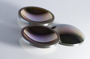
Zirconia (Y-TZP) implants: Excellent results. Zirconia is a ceramic — diamond wire is the natural tool. Surface finish after cutting is comparable to polished ceramic. See our detailed guide on corte de cerámica de circonio for parameter reference.
Cobalt-chrome alloys: More challenging. CoCr is harder and more abrasive than titanium, and wire wear is noticeably higher. Possible, but plan for shorter wire service life and slower feed rates.
PEEK implants with composite reinforcement: Very suitable. PEEK cuts easily, and the low cutting force is ideal for preserving the PEEK-bone interface in osteointegration studies.
The one scenario where endless wire cutting struggles is very large metal cross-sections — an implant that’s 15+ mm in diameter means long wire contact time with metal alone, which accelerates wear. For standard-sized dental or orthopedic implants embedded in bone, this isn’t a problem.
Machine Selection: Which Model to Use for Bone Implant Histological Sections
For histological sample preparation, the workpiece sizes are typically small — most bone-implant samples fit within a 150 × 150 × 150 mm envelope after embedding.
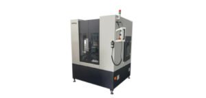
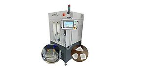
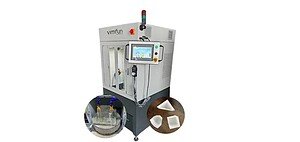
En Vimfun SG15 is a desktop gantry model designed for exactly this scale. It accommodates samples up to 150 × 150 × 150 mm, uses 0.35–0.5 mm wire, and fits on a standard lab bench. Power requirement is 220–240V single phase — no special electrical installation needed.

For larger research samples or multiple simultaneous preparations, the SG20 extends the working envelope to 200 × 200 × 200 mm while maintaining the same desktop form factor and precision characteristics.
Both models use servo-controlled feed axes with ±0.03 mm positioning accuracy. Section thickness repeatability across a series of cuts — assuming the sample is properly mounted and the embedding resin is uniform — is typically within ±0.05 mm, which is adequate for most histological preparation workflows.
The integrated oil mist recovery system handles white mineral oil coolant without requiring external ventilation connections. The machine needs only a power connection to operate — relevant for hospital research labs where installation complexity matters.
What Endless Wire Cutting Does Not Replace
This technology is not a substitute for a precision sectioning saw (IsoMet-type) when you need sections below 100 µm. Wire kerf of ~0.4 mm and the flexibility of the wire make it unsuitable for ultra-thin section production.
It also doesn’t replace microtome-based methods for soft tissue. The diamond wire requires the sample to have sufficient mechanical rigidity — either intrinsic (bone, ceramic, metal) or provided by resin embedding. Soft tissue alone, even when fixed, won’t hold up to wire cutting without a rigid matrix.
For the specific case where you need to cut bone implant histological sections in the 200 µm–2 mm range — where the goal is preserving interface structure rather than achieving optical-grade thinness — endless wire cutting is the right tool. For a deeper look at how wire parameters affect subsurface damage in brittle materials, see Minimizing Subsurface Damage in Diamond Wire Cutting.
Practical Steps to Cut Bone Implant Histological Sections With a Wire Saw
- Prepare a representative sample first. Don’t use your most valuable specimen for a first test. Use a sample with similar material composition — same implant material, similar bone density, same embedding resin.
- Start slow. Begin with a feed rate of 0.5 mm/min and wire speed around 20–25 m/s. Once you confirm the interface is being preserved, you can gradually increase feed rate to optimize throughput.
- Use white mineral oil as coolant. Water-based coolants work for some materials, but for PMMA-embedded bone samples, mineral oil provides better lubrication without the risk of resin swelling or softening that can occur with water over longer cuts.
- Check wire tension after the first few cuts. Tension affects both cutting speed and surface quality. The recommended starting point for 0.35 mm wire is 110–120 N. If you see wire bowing during the cut (visible deflection from straight), increase tension slightly — 5–10 N at a time.
- Evaluate the interface under the microscope before committing to a protocol. Even a single test section tells you a lot about displacement and surface damage. Don’t optimize parameters based on cutting speed alone — the section quality under the microscope is the actual output you’re optimizing for.
Vimfun offers a free sample cutting test — send a representative specimen and the team will return photographs and measurements of the cut result before you purchase equipment. Contact levy@endlesswiresaw.com with your sample description to arrange this.
Resumen
The core problem when you cut bone implant histological sections with conventional saws is not tool quality — it’s cutting mechanism. Tooth-based and blade-based methods generate impact forces and lateral stress that the partially osseointegrated bone-implant interface simply can’t withstand.
Endless diamond wire cutting addresses this at the mechanism level — by replacing impact-based cutting with low-force abrasion. The result is preserved interface structure, higher section yield, and less material removal per cut.
The parameters aren’t complicated. The machines are lab-scale. The main trade-off is cutting speed, which is slow by industrial standards but acceptable for research volumes.
If your current section yield on bone-implant samples is below 70%, this is worth testing before your next sample set.
→ Request a free sample cutting test or get a quotation for the SG15